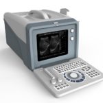
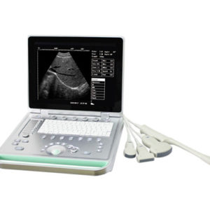
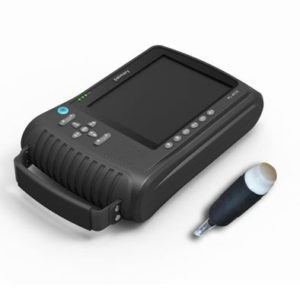
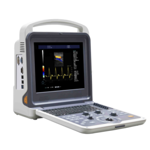
Product Description

Main functions:
Display mode: B, B/B, 4B, B+M, and M
Magnification: ×0.8, ×1.0, ×1.2 (Depth hoist display), and × 1.5,×1 .8, ×2.0 (Depth hoist display)
Dynamic range: 64~96dB adjustable
Focusing: Four sections of dynamic electronic focusing may be elected.
Pre-processes: changeable aperture, dynamic changes of the marks, dynamic filter and edge enhancement, etc.
Pro-processes: 8 kind of γadjustments,16 kinds of color process, line correlation, frame correlation and linear interpolation, etc.
Frequency conversion: 2.5MHz/3.0MHz/3.5MHz/4.0MHz/6.5MHz/7MHz(Matching 6.5MHZ cavity probe and 7 MHz High-frequency linear array probe).
Calculation: distance, perimeter, area, heart rate, pregnancy week (BPD, GS, CRL, FL, AC) and anticipated delivery date.
Note: name, serial numbers of case history, gender, age, 16 body marks (with probe), full-screen character, real-time clock
Puncture guide: The puncture guide line can be displayed under B mode.
Gain control: The total gain, near field and far field can be adjusted successively.
Image polarity: left/right, black/white and up/down reverse.
Fractionated gain: 2 time of enlargements
Movie memory: 256 pictures can be memorized successively when the real-time is displayed.
Image memory: 128 pictures Permanent storage.
Image review: Images can be reviewed successively and checked one by one.
Output interface: 2 groups of SVGA video output may mate with SVGA color monitor. And 2 groups of PAL video output may mate with PAL monitor, video image recording instrument and image workshop, etc. 1 group of USB Connection (Optional).
Main technical index:
Probe: 80Array element R60
Probe frequency: 3.5MHz
Scanning depth: ≥180mm
Lateral resolution: ≤2mm(depth≤80mm), ≤3mm(80< depth≤130mm)
Axial: ≤1mm(depth≤80mm), ≤2mm(80< depth≤130mm)
Dead zone: ≤3 mm。
Geometry position precision: lateral≤5%, axial≤5%
Monitor: 10 inch SVGA high resolution monitor(may choose PAL monitor)
Frames cine loops:256
Power supply scope: AC 220V±22V 50Hz
Input: ≤ 300VA
Successive working hours:≥8h
G/W: 10KGs
N/W: 7KGs
Packing size: 480x380x410mm


Reviews
There are no reviews yet.