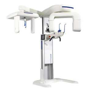
Product Description
Technical Parameters
Scanning system
(1). Type of detector: amorphous silicon flat-panel detectors
(2). Size of detector: 25 x 20cm
(3). Gray scale: 16 bit
(4). Pixel size: 127μm
(5). Resolution ratio: 3.94lp/mm
(6). SFOV: Adjustable, large view Ø 20×17 cm, high definition view Ø 12×10 cm.
X-ray tube and HVG (high-volt generator)
(8). Type of tube: rotating anode.
(9). X-ray beam: Cone-beam, size of beam would change according to the view.
(10). Focus of tube: 0.3mm
(11). Power of tube: 1500W
(12). Current in tube: 2.8mA~9mA
(13). Voltage on tube: 80kV-110kV
(14). HVG: DC
Scanning parameter and image reconstruction
(15). Time of scanning: no more than 18s
(16). Thickness of a single layer: large view-0.2mm, high definition- no more than 0.1mm
(17). Time of rebuilding: large view imaging: no more than 25s, high definition imaging: no more than 60s
(18). Range of rebuilding: 360°
(19). Victor of data transmission: 20Mb/s
Workstation for image:
(20). CPU: Intel E5-1620 3.6GHz
(21). Memory: 8GB DDR3 800MHz ECC
(22). Hard disk: 1TG SATA 3.0Gb/s 7200rpm
(23). Image display matrix: CBCT dedicated 21.5 inch LED backlight IPS displayer
(24). Video memory: GeForce GTX670
(25). OS: Windows 7(64bits) Professional
Functions of the program:
(26).Build new record for patients.
(27). Function of searching for information of patient and saved images
(28). Measurement: distance between two points, continuous distance, angle, user calibration
(29). Display coronal section, sagittal section, axial section and 3D image at one screen
(30).local 360° continuously slicing display
(31). Post processing for multiple image: various plane, vertical plane
(32). Comprehensive diagnosis and lateral projection of cranium
(33). Image processing (adjust luminance , contrast ratio and gamma ray value, and sharpen)
(34). Rebuild 3D image (restore outline of soft tissue)
(35). High definition three-dimensional rebuilding
(36). Burn data disk
(37). Positive for DICOM3.0 Laser Printer print typesetting: multiple saved template, freely composing is also allowed.
(38). Positive for patient’s template
(39). Positive for STL implant template data output
(40). Match the actual need of multiple departments like planting, orthodontics, repairing dental pulp and tooth, maxillofacial, periodontal and so on.
(41). Connect PACS system of hospital, could also accomplish freely transmission in local area network, no limitation for the number of terminal.
(42). Provide image analysis program for free
(43). Compatible for orthodontics, orthognathous, panting and 3D rebuilding professional programs.
(44). Users could choose to install Simplant Pro (Professional planting program) and Simplant O&O (Orthodontics and orthognathous program)
After-sales service
(45). Manufacturer provide on-site repair for 2 years, service aim: “Respond in 2 hours, work out the solution in 8 hours, arrive the scene in 24 hours.”
(46). Provide field technical training
(47). Specification (Chinese and English)
(48). One suit of professional protection suit (Hat, nickerchief and protective clothing)

Reviews
There are no reviews yet.